+84 90 189 0068
 info@dxpsoftware.com

 +84 90 189 0068
 info@dxpsoftware.com
AI-Powered Imaging for Cancer Detection
AI that helps clinicians see what deserves a second look. OncoDx analyzes medical images to highlight suspicious findings and support informed clinical decisions.




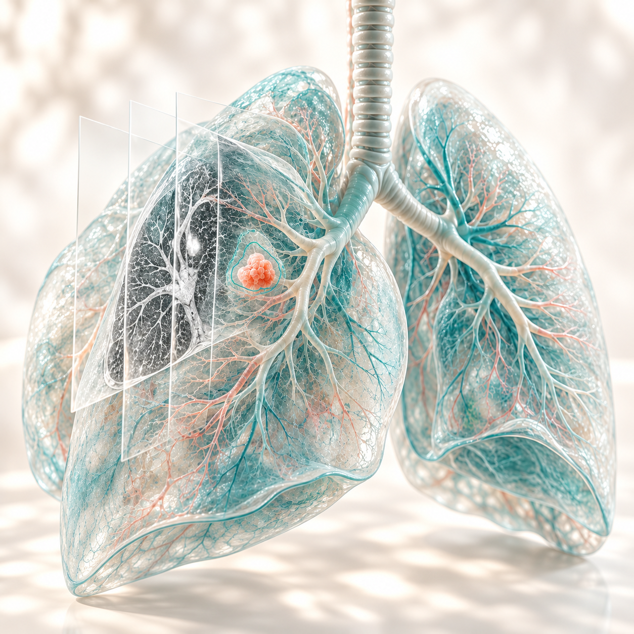
How AI Interprets Medical Images
Medical images contain patterns in tissue structure, shape and appearance. Some findings can be subtle or difficult to distinguish from surrounding anatomy.
OncoDx uses deep learning models trained for specific imaging tasks to identify suspicious regions. Depending on the application, it analyzes CT volumes across multiple slices or processes endoscopy images and video to detect potential lesions.
The findings are presented as visual markers or segmentation overlays, giving clinicians additional information to review alongside the original images and the patient’s clinical context.
From Medical Images to Clinical Review
Hundreds of CT slices. Continuous endoscopy footage. Subtle findings within complex anatomy. OncoDx adds a focused layer of AI analysis, helping clinicians identify areas that warrant closer review.
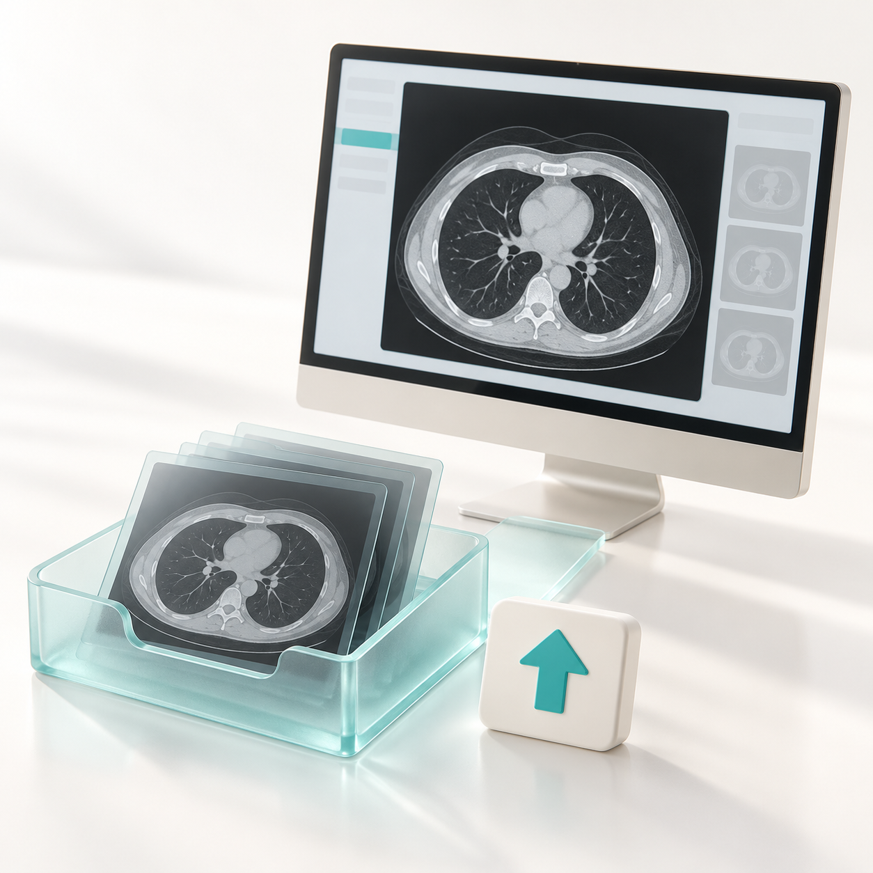
1. Image input
Upload a supported CT study, endoscopy image or video, or receive input through a configured video connection.
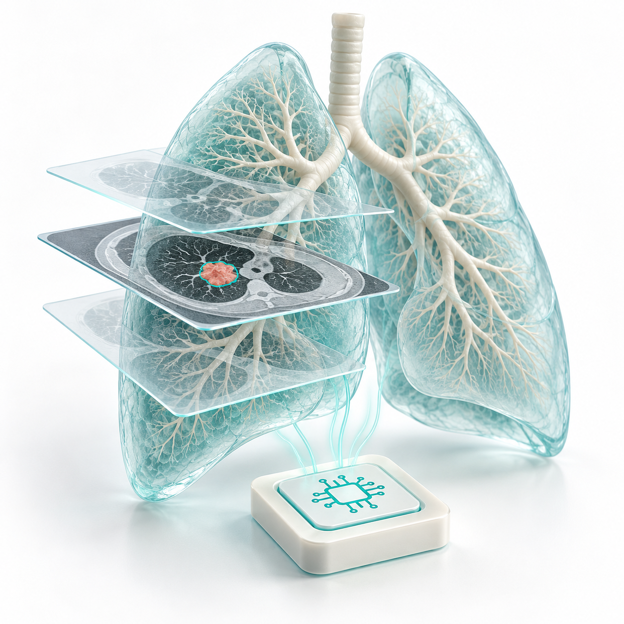
2. AI analysis
A specialized AI model analyzes the images and marks suspected regions for closer inspection.
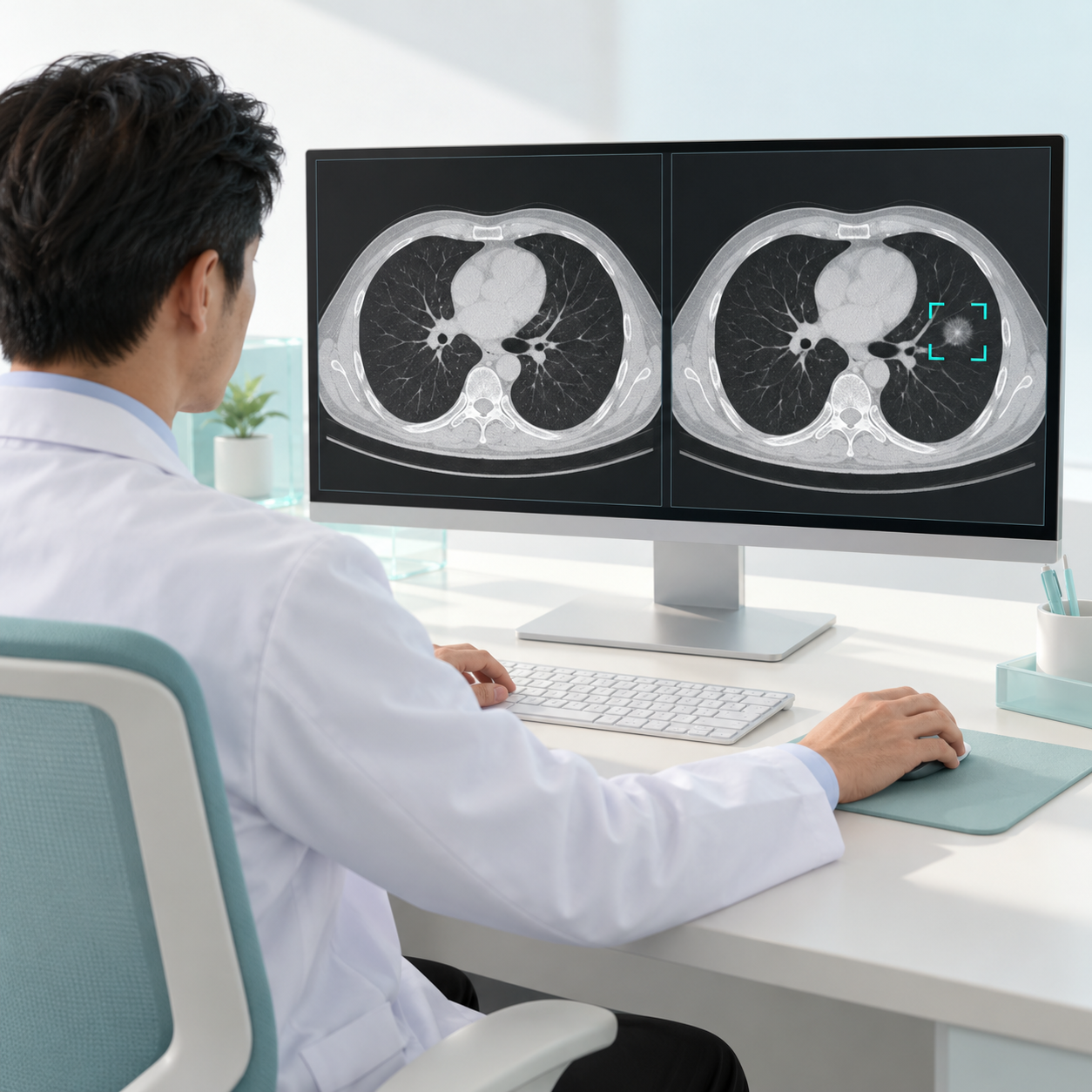
3. Clinical review
The clinician examines AI findings alongside the original images and clinical information to determine the next step.
Four cancer applications.
A growing field of vision.
Different organs. Different imaging challenges. Dedicated models developed for the anatomy and task at hand.
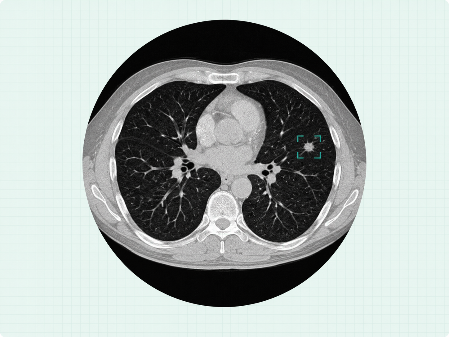 CHEST CT / RESEARCH EXAMPLE
A second look at pulmonary nodules.
Analyze the full CT volume to flag suspected pulmonary nodules for radiologist review.
- DICOM series input and volumetric 3D analysis
- Localized regions with model confidence scores
- Lung-field filtering to reduce out-of-lung predictions
A detected nodule is not a diagnosis of lung cancer.
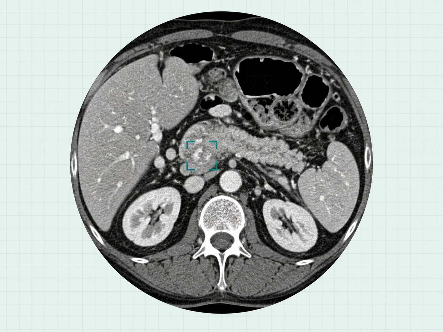 PANCREATIC ANATOMY / ILLUSTRATION
See the pancreas in context.
A multi-model pipeline combines pancreatic anatomy, suspected lesions and duct morphology to support PDAC assessment.
- Pancreas and suspected lesion segmentation
- Pancreatic duct analysis and morphological features
- Combined features for case-level PDAC classification
Focused on pancreatic ductal adenocarcinoma (PDAC).
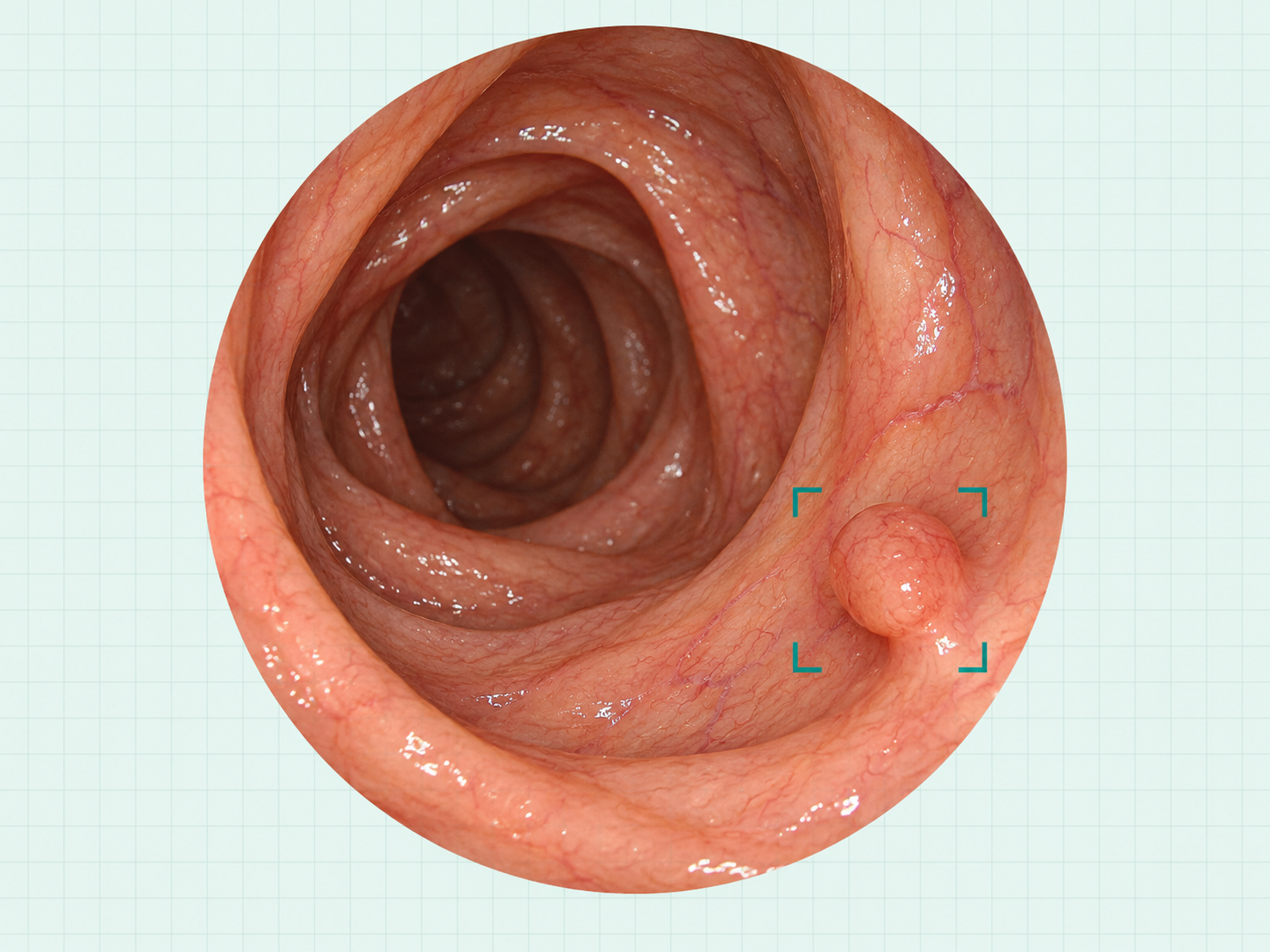 COLORECTAL ANATOMY / ILLUSTRATION
Keep attention on the polyp.
Analyze colorectal endoscopy images and video to highlight suspected polyps during visual review.
- Polyp detection and bounding-box localization
- Model confidence displayed with detected regions
- Video processing optimized for responsive visual support
Polyp detection does not determine benign or malignant pathology.
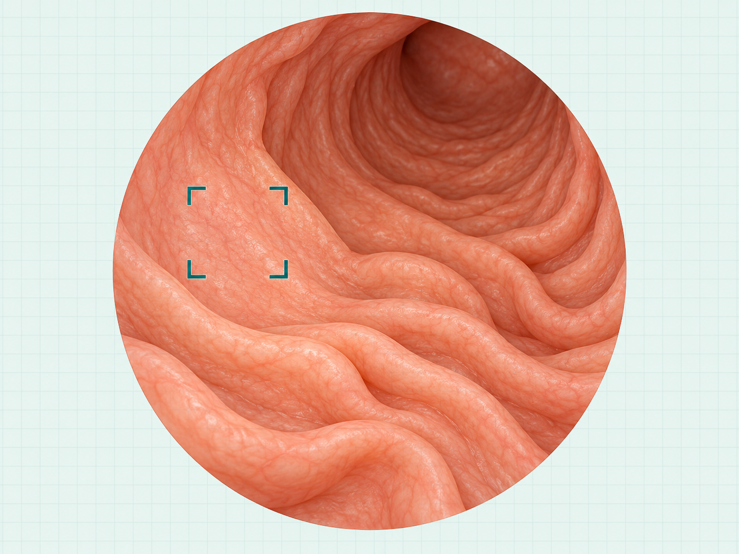 GASTRIC ANATOMY / ILLUSTRATION
A broader view of gastric imaging.
Gastric cancer is one of the four OncoDx application areas, focused on supporting review of suspicious regions.
- A dedicated focus within the OncoDx portfolio
- Application scope defined by model version
- Contact DXP for current capabilities and evaluation options
Detailed input specifications and performance are not yet published here.

“Our goal with OncoDx is to help clinicians detect even small nodules and subtle abnormalities. We combine AI research with specialized medical image analysis to make suspicious findings easier to locate and review, supporting informed clinical judgment and guiding further assessment.”
Dr. Nguyen Duc Phuong CEO & Head of AI R&D LabExplore OncoDx in Action
Discover how AI highlights suspicious findings in medical images. Book a demo